February 23, 2026
A communication guide for dentists
Why Patients Often Question the Safety of Dental Imaging
Many patients hesitate the moment imaging is mentioned. They may associate the word radiation with risk, or they may simply not know how modern digital systems differ from the equipment used in the past. This uncertainty can make simple conversations feel more complex than they need to be.
As clinicians, we work with digital imaging every day and understand its value, clarity, and safety. Patients do not have that perspective. Clear explanations can transform a moment of concern into an opportunity for reassurance and trust.
This guide supports dentists in framing imaging discussions in a way that helps patients understand why it is safe, why it is used, and how it supports their oral health.
How to Explain the Safety of Modern Dental Imaging
Digital dental imaging uses advanced systems designed to reduce dose, focus the beam, and capture information quickly. Patients do not need technical detail. They need simple, relatable points that make sense to them.
Key messages to share with patients
• The radiation dose is very low and highly targeted
• Digital sensors capture images instantly which reduces exposure
• The equipment focuses on specific teeth rather than the entire head
• Protective aprons and collars may be used where appropriate
• Imaging is only taken when clinically needed
• Your practice follows strict safety protocols every time imaging is completed
These points help patients understand that modern imaging is built around safety, precision, and efficiency.
Using Everyday Comparisons to Reassure Patients
Most patients have no reference point for what a low dose actually means. Comparing dental imaging with ordinary life experiences helps remove anxiety and gives them a clear sense of perspective.
Everyday Radiation Comparison
| Activity or Exposure | Relative Level | How to Communicate It to Patients |
| A single dental X ray | Very low | One of the lowest levels used in healthcare |
| A full mouth series | Low | Still far below many medical scans |
| A day of natural background radiation | Low to moderate | Something everyone is exposed to daily |
| A long flight | Low to moderate | Often higher than a dental X ray because of altitude |
| Medical CT scan | High | Demonstrates how low dental imaging is by comparison |
Patients often relax when they understand dental imaging sits at the lowest end of the scale.
Helping Patients Understand Why Imaging Is Recommended
When patients know why a particular view is needed, trust increases. Instead of simply saying an X ray is required, explain what the image helps you assess and how it improves the accuracy of their care.
Common Imaging Types and How to Describe Them to Patients
| Imaging Type | What It Shows | Suggested Patient Explanation |
| Bitewing | Early decay and bone levels | Helps us check between teeth where we cannot see visually |
| Periapical | Roots and surrounding bone | Helps us find infections or cracks below the surface |
| Panoramic OPG | Jaws, joints, wisdom teeth | Gives a wide overview of your whole mouth |
| Cephalometric | Jaw position and growth | Used for orthodontic planning and monitoring |
| CBCT | Three dimensional structures | Helps us plan complex treatment safely and accurately |
Framing each imaging type in practical, everyday language makes recommendations easier for patients to understand and accept.
How to Communicate Radiation Safety Protocols
Patients feel reassured when they know safety is actively managed. A short explanation of your clinic’s protocol can help reduce hesitation.
What to tell patients
• Our systems are modern digital units designed for very low dose imaging
• We use protective equipment when it is appropriate
• Images are taken only when they are clinically justified
• The beam is tightly focused to reduce exposure
• Our team is trained to follow strict regulatory protocols
Concise statements help patients recognise that imaging is deliberate, controlled, and necessary for their care.
Explaining the Role of Imaging in Oral Health
Many dental conditions develop quietly. Patients often assume that no pain means no problem. Clarifying how imaging supports prevention can reshape their understanding.
Situations you can explain to patients
• First visits where a baseline is needed
• Monitoring bone health around teeth
• Checking for decay that cannot be seen visually
• Assessing wisdom teeth or jaw development
• Investigating pain or suspected infection
• Planning for restorative, surgical, or orthodontic treatment
When patients see imaging as a tool that prevents issues from becoming more complex, they feel more comfortable about receiving it.
How the Right Equipment Partner Supports Safe Conversations
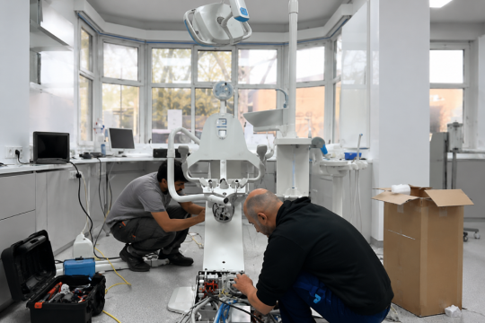
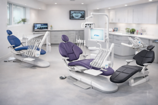
Dentists can reassure patients more confidently when equipment consistently delivers clear images and supports low dose protocols. Medical Dental Solutions plays an important role in this by supplying and maintaining digital imaging systems, sensors, consumables, chairs, and surgery equipment for dental practices across Australia.
Reliable equipment helps clinicians deliver safe, predictable imaging every day and strengthens patient confidence during discussions.
Frequently Asked Questions You May Receive from Patients
These responses can be adapted for chairside conversations.
1. Are dental X rays safe for my family and me
Yes. Modern digital imaging uses low radiation technology with very targeted fields designed to minimise exposure.
2. How often will I need imaging
Only when it is clinically required. The frequency depends on your individual oral health and treatment needs.
3. Is imaging safe during pregnancy
Imaging is usually postponed unless absolutely necessary. If required, safety precautions are used to minimise exposure.
4. Why do I need an X ray if nothing hurts
Many dental problems do not cause pain until they are advanced. Imaging helps detect issues early.
5. Do digital X rays use less radiation
Yes. Digital imaging requires significantly less exposure than older film based systems.
6. Can I choose not to have imaging
You can always discuss questions or concerns. Your dentist will explain why imaging is recommended and how it supports your care.