December 14, 2024
Dental care has come a long way, with advancements in technology playing a crucial role in improving diagnostic accuracy and treatment outcomes. One of the key innovations in dentistry is dental imaging. This technique has become an indispensable tool for dental professionals, enabling them to detect problems early, plan treatments more accurately, and ensure patient safety throughout their care journey.
So, why is dental imaging so vital, and how does it benefit you as a patient? Let’s delve into the importance of dental imaging in modern dental care.
What is Dental Imaging and Why is it Essential?
Dental imaging refers to the use of advanced technology to capture detailed images of the teeth, gums, and jawbone. These images help dentists diagnose oral health issues that might not be visible during a routine dental exam. Using digital X-rays, Cone Beam CT scans, panoramic X-rays, and intraoral cameras, dental professionals can detect problems such as cavities, gum disease, infections, tooth fractures, and even early signs of oral cancer.
How Does Dental Imaging Work?
Dental imaging technology works by capturing high-resolution images of the oral structures. Modern methods, such as digital X-rays, are far more efficient and safer than older methods, using up to 80% less radiation. The images are often displayed on a screen for the dentist to evaluate immediately, allowing them to make quick and accurate decisions about your care.
The digital X-ray technique is one of the most commonly used methods in dental imaging. The process involves using a small digital sensor placed inside the mouth to capture images of the teeth and surrounding tissues. The results are sent to a computer, where the dentist can quickly assess them.
Cone Beam CT (CBCT), another popular dental imaging method, provides a 3D view of the teeth, gums, and bone structure. This detailed imaging technique is essential for complex treatments like dental implants, as it allows for precise planning and accurate placement.
What are the Benefits of Dental Imaging?
Dental imaging offers a wide range of benefits that significantly improve the quality of care provided. Let’s look at some of the key advantages:
Early Detection of Dental Issues
The primary benefit of dental imaging is its ability to detect dental issues in their earliest stages. For example, cavities that are not visible during a routine dental check-up can be spotted early using X-rays, allowing for more conservative and less invasive treatment. Similarly, gum disease, infections, and abscesses can be identified before they escalate into more serious problems.
Accurate Treatment Planning
Dental imaging provides a detailed and comprehensive view of the mouth, jaw, and teeth, which helps dental professionals develop more effective treatment plans. Whether it’s planning for dental implants, orthodontics, or root canal treatments, having accurate images ensures that the dentist can approach each case with precision.
Minimised Radiation Exposure
Unlike traditional X-rays, modern digital dental imaging systems use significantly less radiation. The reduced radiation exposure is a huge advantage for both patients and dental practitioners, ensuring that diagnostic procedures are safer while still providing high-quality images.
Enhanced Patient Education
One of the most significant advantages of dental imaging is its role in patient education. Using intraoral cameras, patients can visually see their own dental problems, which helps them better understand the need for certain treatments. This can lead to greater patient compliance and a more collaborative approach to dental health.
Better Diagnosis and Monitoring
Dental imaging enables precise diagnosis and monitoring of dental conditions over time. For example, patients with gum disease can have their condition tracked through regular X-rays, allowing for the early detection of bone loss or other complications. This leads to more targeted interventions and ultimately better oral health outcomes.
How Does Dental Imaging Improve Preventative Care?
The importance of dental imaging is not just about diagnosing existing issues; it’s also a crucial tool for preventative care. With detailed imaging, dentists can assess a patient’s oral health on an ongoing basis and catch potential issues before they become significant problems.
For instance, regular dental X-rays can help detect early signs of tooth decay or gums recession, allowing for interventions that prevent further damage. This proactive approach helps reduce the need for costly and invasive treatments down the line. By identifying problems early, dental imaging helps save both time and money for patients.
What Are the Different Types of Dental Imaging?
Dental professionals have access to a variety of imaging techniques, each offering specific benefits depending on the nature of the issue being investigated. Below are the most commonly used types of dental imaging:
| Type of Dental Imaging | Description |
| Intraoral X-rays | Provides clear images of individual teeth and the surrounding bone. Ideal for detecting cavities, bone loss, and infections. |
| Panoramic X-rays | Captures the entire mouth in one image, including the teeth, upper and lower jaws, and surrounding tissues. Used to assess overall dental health and plan treatments like extractions or orthodontics. |
| Cone Beam CT (CBCT) | A 3D imaging technique that gives a comprehensive view of the teeth, jaw, and surrounding structures. Essential for planning dental implants and complex oral surgeries. |
| Digital X-rays | An advanced form of traditional X-rays that use digital sensors for faster, more detailed imaging. Uses less radiation and offers immediate results. |
| MRI (Magnetic Resonance Imaging) | Primarily used for soft tissue evaluation, though rarely used in dentistry. Occasionally used in diagnosing temporomandibular joint (TMJ) disorders or other soft tissue issues in the jaw. |
| Intraoral Cameras | Small cameras used to capture detailed images of the inside of the mouth, offering real-time visuals of issues like cavities, gum disease, and cracks in teeth. |
What Are the Risks of Dental Imaging?
While dental imaging is safe, there are some risks associated with it. The primary concern is radiation exposure, though this is minimal with modern digital systems. Dentists take every precaution to minimise exposure, using protective lead aprons and limiting the number of X-rays taken.
It is important to inform your dentist of any potential risks, such as pregnancy, which may require extra precautions or the avoidance of certain imaging techniques altogether.
How Does Dental Imaging Help in Complex Procedures?
Dental imaging is indispensable in more complex dental treatments, such as dental implants or root canal therapy. By using advanced imaging systems like Cone Beam CT, dentists can precisely evaluate bone structure and ensure the correct placement of dental implants.
For root canal treatments, dental X-rays help determine the extent of infection or damage within the tooth, guiding the dentist throughout the procedure. Without this information, treatments could be less effective or more invasive than necessary.
What Role Does Dental Imaging Play in Orthodontics?
In orthodontics, dental imaging plays a critical role in developing personalised treatment plans. Panoramic X-rays provide a complete view of the teeth and jaw, allowing orthodontists to assess the alignment of teeth, the health of the jawbone, and plan for braces or aligners.
Using digital imaging, orthodontists can track the movement of teeth during treatment, ensuring the procedure stays on course and delivers the best possible results.
How Can Dental Imaging Improve Your Oral Health?
Dental imaging allows for the early detection and intervention of many oral health issues, from cavities and gum disease to more complex conditions like tooth abscesses or impacted teeth. The precise diagnostic capabilities provided by modern imaging ensure that you receive timely, effective treatment, helping you maintain your oral health for years to come.
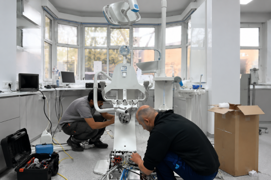
If you are looking to enhance your dental care with the latest technology, consider consulting Medical Dental Solutions. We offer state-of-the-art solutions designed to improve your practice’s diagnostic accuracy and patient care experience. With our help, your dental practice can provide a higher standard of care, leading to better patient outcomes and satisfaction.
References:
- Australian Dental Association. (2020). Dental Radiography – Safety and Procedures. ADA
- Australian Society of Dental Implants. (2021). Cone Beam CT in Implantology. ASDI
Frequently Asked Questions
1. How often should I have dental imaging done?
Typically, dental imaging is done once a year during your routine check-up, or more frequently if you have a history of dental issues or are undergoing certain treatments like braces or implants.
2. Is dental imaging safe?
Yes, modern dental imaging techniques, particularly digital X-rays, use significantly lower radiation than older methods. Protective measures such as lead aprons are also used to minimise exposure.
3. What is Cone Beam CT and why is it used?
Cone Beam CT provides a 3D image of the mouth, jaw, and surrounding areas. It is especially useful in complex procedures such as dental implants, where precise planning is crucial for success.
4. Does dental imaging hurt?
No, dental imaging is a painless and non-invasive procedure. Digital X-rays and CBCT scans are typically quick and comfortable for the patient.
5. Can dental imaging detect oral cancer?
While dental imaging can identify abnormal growths or lesions, further testing may be required to definitively diagnose oral cancer.
6. How does dental imaging help in orthodontics?
In orthodontics, dental imaging helps to assess the alignment of teeth, the health of the jawbone, and plan for braces or clear aligners, ensuring a personalised treatment plan that delivers the best results.